Introduction
Near-infrared spectroscopy (NIRS) is a non-invasive neuroimaging technique that recently has been developed to measure the changes of cerebral blood oxygenation associated with brain activities. To date, for functional brain mapping applications, there is no standard on-line method for analyzing NIRS data. In this work, a novel on-line NIRS data analysis framework taking advantages of both the general linear model (GLM) and the Kalman estimator is devised. The Kalman estimator is used to update the GLM coefficients recursively, and one critical coefficient regarding brain activities is then passed to a t-statistical test. The t-statistical test result is used to update a topographic brain activation map. Meanwhile, a set of high-pass filters is plugged into the GLM to prevent very low-frequency noises, and an autoregressive (AR) model is used to prevent the temporal correlation caused by physiological noises in NIRS time series. A set of data recorded in finger tapping experiments is studied using the proposed framework. The obtained results suggest that the method can effectively track the task related brain activation areas, and prevent the noise distortion in the estimation while the experiment is running. Thereby, the potential of the proposed method for real-time NIRS-based brain imaging was demonstrated. This work presents a novel on-line approach for analyzing NIRS data for functional brain mapping applications. This approach demonstrates the potential of a real-time-updating topographic brain activation map.
Detection of event-related hemodynamic response
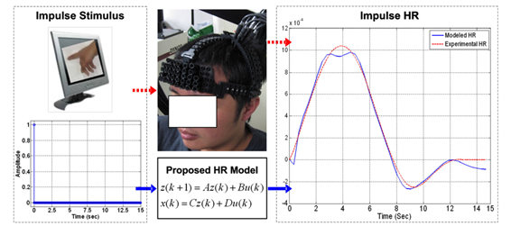
In this lab, research is done on state-space hemodynamic model by which any event-related hemodynamic prediction function (i.e., the basis function of the design matrix in the general linear model) is obtained as an output of the model. To model the actual event-related behavior during a task period (intra-activity dynamics) besides the contrasting behavior among the different task periods and against the rest periods (inter-activity dynamics), the modular system is investigated by parametric subspace-based state-space modeling of actual hemodynamic response to an impulse stimulus. This model provides a simple and computationally efficient way to generate the event-related basis function for an experiment by just convolving the developed hemodynamic model with the impulse approximation of the experimental stimuli.
[Comparison of two hemodynamic responses]
The demonstration of the stated findings is carried out by conducting finger-related experiments with slow- and fast-sampling near-infrared spectroscopy instruments to model and validate the cortical hemodynamic responses.
[Brain activation maps drawn using right index finger tapping]
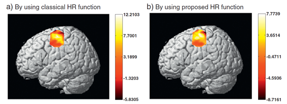
The generated basis functions of the finger-related experiments are adapted from real data to validate the incorporation of non-delayed and real-time event-related features and to effectively demonstrate a dynamic-modeling-based online framework. The proposed method demonstrates potential in estimating event-related intra- and inter-activation dynamics and thereby outperforms the classical Gaussian approximation method.
[Results of the multi-cortex finger grasping experiment]
For more details, see “Detection of Event-Related Hemodynamic Response to Neuroactivation by Dynamic Modeling of Brain Activity,” Neuroimage, Vol. 63, No 1, pp. 553-568, October 2012.
Trial-to-trial variability and functional connectivity
The reduction of trial-to-trial variability (TTV) in task-evoked functional near-infrared spectroscopy signals by considering the correlated low-frequency spontaneous fluctuations that account for the resting-state functional connectivity in the brain is investigated. A resting-state session followed by a task-state session of a right hand finger tapping task has been performed on five subjects.
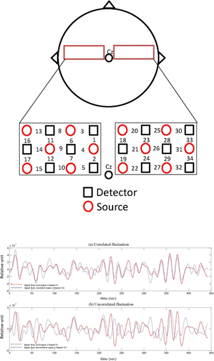
[Optode position and raw fNIRS signal]
Significant ipsilateral and bilateral resting-state functional connectivity has been detected at the subjects motor cortex using the seed correlation method. The correlation coefficients obtained during the resting-state are used to reduce the TTV in the signals measured during the task sessions. The results suggest that correlated spontaneous low-frequency fluctuations contribute significantly to the TTV in the task evoked fNIRS signals.
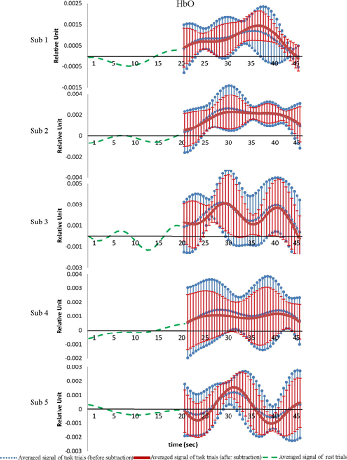
[HBO signals for five subjects]
For more details, see "Reduction of Trial-to-Trial Variations in Functional Near-Infrared Spectroscopy Signals by Accounting for Resting-State Functional Connectivity," Journal of Biomedical Optics, Vol. 18, No. 1, Article ID: 017003, January 2013.
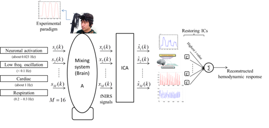
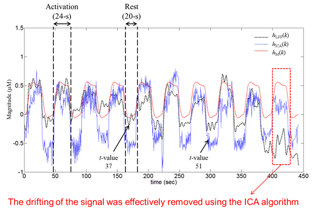
Noise reduction in fNIRS signals using independent component analysis
In this study, we investigate the independent component analysis (ICA) method as a means of analyzing fNIRS signals and removing noise. The proposed ICA method could effectively separate physiological noises (spontaneous fluctuation and respiratory signals) and motion artifacts for all channels employed.
[The proposed ICA method]
After reconstruction of the independent components, significant t-value increases for all subjects, in a comparative evaluation with conventional low-pass filtering. Overall, the results show the applicability of the ICA-based method to noise-contamination reduction in brain mapping.
[Oxy-hemoglobin comparison from representative subjects]
For more details, see “Noise reduction in functional near-infrared spectroscopy signals by independent component analysis,” Review of Scientific Instruments, Vol. 84, No. 7, AN: 073106,July 2013.
State-space approach
This work presents state space models of the hemodynamic response (HR) of fNIRS to an impulse stimulus in three brain regions: motor cortex (MC), somatosensory cortex (SC), and visual cortex (VC). Three impulse HRs experimentally obtained from nineteen healthy subjects were averaged. The averaged signal was converted to state space equation using the subspace method. The advantage of the proposed method is its easy applicability in generating the expected HR to arbitrary stimuli in an online (or real-time) imaging.
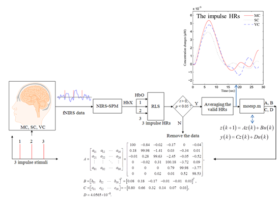
[The scheme for reconstruction of impulse HRs in three brain regions using state-space approach]
The HR to an impulse stimulus in different brain regions (i.e., MC, SC, and VC) was investigated. Our results show that the activation peak and undershoot-peak of HbO in MC are noticeably higher than those in SC and VC. The time-to-peak in three brain regions are almost the same. The time-to-undershoot peak in VC is largest among three. The HbO decreases in the early stage (~0.46 s) in MC and VC, but not in SC. In each cortex, an impulse HR function in state-space form is first developed. Then, it is used to generate the expected HR for arbitrary stimuli. It is noteworthy that the differences of measured impulse HRs in three brain regions were fully integrated in the state space equations while the canonical HR function in the form of double-gamma function cannot.
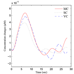
[Comparison of the impulse HbOs in three br ain regions]
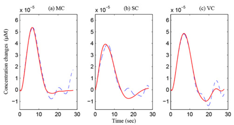
[Comparison between the experimental HbOs (blue dashed curves) and the reconstructed ones (red solid curves): (a) MC, (b) SC, and (c) VC]
For more details, see “State-space models of inpulse hemodynamic responses over motor, somatosensory, and visual cortices,” Biomedical Opitcs Express , Vol. 5, No. 6, pp. 1778-1798 , June 2014.
Music processing with noises in human auditory cortex
The present study is to determine the effects of background noise on the hemispheric lateralization in music processing to four different auditory environment: music segments only, noise segments only, music + noise segments, and the entire music interfered by noise segments.
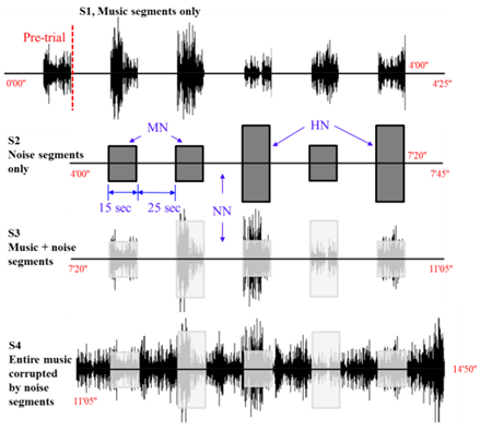
[Experimental paradigm: Four different sound conditions]
The difference between the mean and the minimum value of a given hemodynamic response has been used as a feature to classify whether the hemispheric lateralization occurred or not. The main finding of this study is that the background noise affects the hemodynamic response in the activation of the auditory cortex in music processing.
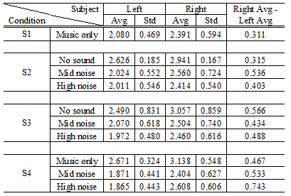
[The difference value: Average of only those cases where Right > Left]
For more details, see "Lateralization of music processing with noises in the auditory cortex: an fNIRS study, " Frontiers in Behavioral Neuroscience, Vol. 418, No. 8, AN: 418 (1-9), December 2014.